The process or technique of producing images of an opaque object on photographic film or on a fluorescent screen by means of radiation (either particles or electromagnetic waves of short wavelength, such as X-rays and gamma-rays). The photograph produced is called a radiograph .
In a medical radiograph part of a patient's body is exposed to X-rays so that a shadow image of that body part is produced on a film placed behind the body. The image is formed by the varying degrees of attenuation offered by the various media within the body. The degree of attenuation depends on three main factors:
1. The attenuating character of the medium. This is usually characterized by an attenuation coefficient .
2. The density of the medium.
3. The thickness of the structure.
Bone offers the greatest degree of attenuation, followed by soft tissue, and then pockets of air. Conventional radiography cannot easily distinguish between different types of soft tissue (e.g. liver and kidney tissues) owing to their similar attenuation characters and densities. Bone, however, is very easily distinguished from neighbouring soft tissue, such as muscle.
If better contrast is required, a contrast medium, such as barium, can be used, which preferentially absorbs X-rays and enables otherwise difficult anatomical features to be observed. Kidney, arterial, and intestinal functions are examined by this method, using various contrast media.
When an X-ray interacts with an atom in the medium, there are several possible mechanisms that may attenuate the X-ray. The attenuation mechanism that the X-ray undergoes depends on its energy. The main attenuation mechanisms are:
Simple scattering: this occurs when the X-ray has insufficient energy to cause an ionization of the atom. The X-ray is simply deflected without significant change of energy.
Photoelectric effect: the X-ray transfers all of its energy to an electron in the atom and therefore ionizes the atom (see photoelectric effect).
Compton scattering: the high-energy X-ray collides with an electron, transferring both energy and momentum to it. The X-ray recoils with a loss of energy (see Compton effect).
Pair production: this mechanism occurs at very high energies. Within the field of the interacting atomic nucleus, the X-ray spontaneously creates a positron–electron pair.
The table below shows the thresholds for the various attenuation mechanisms and their variation with the atomic number Z of the medium material and the energy E of the X-ray.
The optimum X-ray energy for diagnostic radiography is in the range of the photoelectric effect. At about 30 keV, the photoelectric effect dominates the attenuation of the X-radiation and leads to a better contrast between media of different atomic numbers. The Z3 variation of the attenuation within the photoelectric-dominated regime means that the distinction between media of different Z is more pronounced.
| Mechanism | Variation of the attenuation with the energy (E) of the X-ray | Variation of the attenuation with the atomic number (Z) of the medium substance | Energy range in which this kind of attenuation is dominant |
| simple scattering | α 1/E | αZ2 | 1–20 keV |
| photoelectric effect | α 1/E3 | αZ3 | 1–30 keV |
| Compton scattering | falls gradually with E | independent | 30 keV – 20 MeV |
| pair production | rises slowly with E | αZ2 | above 20 MeV |
Air has a higher average Z than soft tissue, which consists of mostly carbon (6C) and hydrogen (1H). However, the low average density of air means that it does not attenuate X-rays strongly. Barium, on the other hand, has an atomic number of 56, and thus attenuates very strongly, which is why it is used as a contrast medium in barium meals and barium enemas.
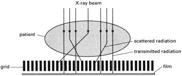
Diagnostic radiography requires high-quality images of internal structures. This means that a high contrast and low blurring must be achieved. It is therefore important to prevent X-rays falling onto the film, which have scattered off many different parts of the body. This is made possible by the introduction of a grid between the patient and the film as shown in the diagram.
The grid consists of narrow lead strips that allow passage of the main unabsorbed beam, but intercept randomly scattered X-rays. The entire grid is oscillated during the formation of the radiograph in order to prevent the formation of its own well-defined X-ray shadow. Before the X-ray beam interacts with the patient's body it is controlled by a diaphragm to limit the beam width. This gives a relatively well-defined beam that can be directed onto the desired area. Wide beams are undesirable because they generate unwanted scattering and deliver an unnecessarily high dose to the patient. Longer exposure at low beam intensity can, in principle, improve the contrast. However, blurring becomes more likely owing to the involuntary movements of the patient during the scan.
A natural magnification of the subject structure occurs during an X-ray scan because the X-radiation source acts as a point source. The area of the X-ray shadow is greater than that occupied by the two-dimensional projection of the structure under examination. In practice the X-ray source is not a point and the image is surrounded by a penumbra, which constitutes a relatively blurred region. The more point-like the source, the smaller the penumbra on the image.
Radiography (medical)
- radiolysis
- radiometric dating
- radionuclide
- radiosonde
- radiotherapy
- radiotransparent
- radium
- radius of curvature
- radius of gyration
- radius vector
- radon
- rainbow
- RAM
- Raman effect
- ramjet
- Ramsay, Sir William
- Ramsden eyepiece
- random alloy
- random walk
- Rankine cycle
- Rankine temperature scale
- Raoult's law
- rarefaction
- raster
- rational number