The absorption of electromagnetic radiation at a suitable precise frequency by a nucleus with a nonzero magnetic moment in an external magnetic field. The phenomenon occurs if the nucleus has nonzero spin, in which case it behaves as a small magnet. In an external magnetic field, the nucleus's magnetic moment vector precesses about the field direction but only certain orientations are allowed by quantum rules. Thus, for hydrogen (spin of ½) there are two possible states in the presence of a field, each with a slightly different energy. Nuclear magnetic resonance is the absorption of radiation at a photon energy equal to the difference between these levels, causing a transition from a lower to a higher energy state. For practical purposes, the difference in energy levels is small and the radiation is in the radiofrequency region of the electromagnetic spectrum. It depends on the field strength.
NMR can be used for the accurate determination of nuclear moments. It can also be used in a sensitive form of magnetometer to measure magnetic fields. In medicine, magnetic resonance imaging (MRI ) has been developed, in which images of tissue are produced by magnetic-resonance techniques.
The main application of NMR is as a technique for chemical analysis and structure determination, known as NMR spectroscopy . It depends on the fact that the electrons in a molecule shield the nucleus to some extent from the field, causing different atoms to absorb at slightly different frequencies (or at slightly different fields for a fixed frequency). Such effects are known as chemical shifts . There are two methods of NMR spectroscopy. In continuous wave (CW ) NMR, the sample is subjected to a strong field, which can be varied in a controlled way over a small region. It is irradiated with radiation at a fixed frequency, and a detector monitors the field at the sample. As the field changes, absorption corresponding to transitions occurs at certain values, and this causes oscillations in the field, which induce a signal in the detector. Fourier transform (FT ) NMR uses a fixed magnetic field and the sample is subjected to a high-intensity pulse of radiation covering a range of frequencies. The signal produced is analysed mathematically to give the NMR spectrum. The most common nucleus studied is 1H. For instance, an NMR spectrum of ethanol (CH3CH2OH) has three peaks in the ratio 3:2:1, corresponding to the three different hydrogen-atom environments. The peaks also have a fine structure caused by interaction between spins in the molecule. Other nuclei can also be used for NMR spectroscopy (e.g. 13C, 14N, 19F) although these generally have lower magnetic moment and natural abundance than hydrogen. See also electron-spin resonance.
MAGNETIC RESONANCE IMAGING
This diagnostic imaging technique is based on nuclear magnetic resonance (NMR), in which protons interact with a strong magnetic field and with radio waves to generate electrical pulses that can be processed in a similar way to computerized tomography. The medical application of NMR began in the 1950s, but the first images of live patients were not produced until the late 1970s. Though expensive, the technique has become a very promising medical tool. Images produced by MRI are similar to those produced by computerized tomography using X-rays, but without the radiation hazard.
A major factor in the high costs of MRI is the need for a superconducting magnet to produce the very strong magnetic fields (0.1–2 tesla). A niobium-titanium alloy, which becomes superconducting at -269°C, is used to construct the field coils. These need to be immersed in liquid helium backed up with liquid nitrogen. Superimposed on this large magnetic field are smaller fields, with known gradients in two directions. These gradient fields produce a unique value of the magnetic field strength at each point within the instrument (see illustration).
Some nuclei in the atoms of a patient's tissues have a spin, which makes them behave as tiny nuclear magnets. The purpose of the large magnetic field is to align these nuclear magnets. Having achieved this alignment, the area under examination is subjected to pulses of radio-frequency (RF) radiation. At a resonant frequency of RF pulses the nuclei under examination undergo Larmor precession. This phenomenon may be thought of as a ‘tipping’ of the nuclear magnets away from the strong field alignment. The nuclear magnets then precess, or ‘wobble’, about the axis of the main field as the nuclei regain their alignment with that field.
The speed at which the nuclei return to the steady state gives rise to two parameters, known as relaxation times . Because these relaxation times for nuclei depend on their atomic environment, they may be used to identify nuclei. Small changes in the magnetic field produced as the nuclei precess induce currents in a receiving coil. These signals are digitized before being stored in a computer.
The resulting set of RF pulse sizes and sequences identify a variety of resonance situations. By analysing these sequences and knowing the unique value of magnetic field strength within the volume under investigation, the resonance signals may be decoded to give estimates of the composition of the patient's tissues. A three-dimensional map of the composition can then be produced, using colour to indicate contrast between differing tissue compositions.
MRI has produced spectacular results in studies of the brain and central nervous system, providing excellent images of delicate structures without the risk of the damage associated with ionizing radiation. Systems using very strong fields, in the region of 2 tesla or above, produce images of extremely high quality.
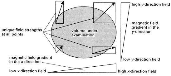
MRI: the way unique field strengths are produced at different points in a specimen.
- Fermi pressure
- Fermi, Enrico
- Fermi-Dirac statistics
- fermion
- fermium
- ferrimagnetism
- ferrite
- ferroalloys
- ferroelectric materials
- ferromagnetism
- fertile material
- FET
- Feynman diagram
- fibre optics
- field
- field coil
- field emission
- field lens
- field magnet
- field-effect transistor
- field-emission microscope
- field-ionization microscope
- file
- film badge
- filter